In the intricate world of auditory processing, the cochlear nerve plays a pivotal role. This article sheds light on the fascinating journey of sound signals from the ear to the brain and explores the connection between the cochlear nerve and the brain. Understanding these intricate processes can enhance our appreciation for the complexity and wonder of the human auditory system. It is important to note that while this article provides valuable insights into the topic, individual cases may vary and it is always recommended to consult with a qualified medical professional for personalized advice and guidance.
Understanding the Cochlear Nerve
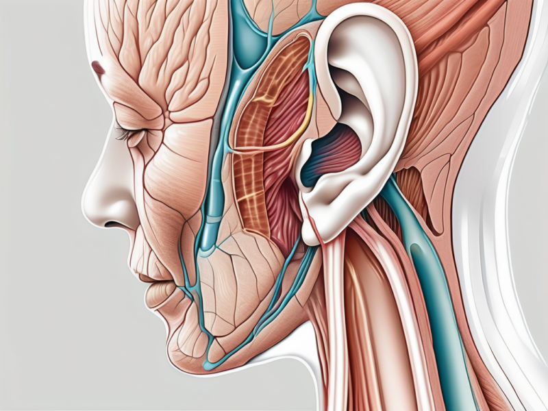
The cochlear nerve is a crucial component of the auditory pathway, responsible for transmitting auditory information from the ear to the brain. It is one of the branches of the vestibulocochlear nerve, also known as the eighth cranial nerve.
Anatomy of the Cochlear Nerve
The cochlear nerve consists of two primary divisions: the cochlear ganglion and the auditory nerve fibers. The cochlear ganglion, located within the inner ear, contains sensory neurons that receive signals from the hair cells in the cochlea. These sensory neurons then send the encoded auditory information to the brain.
The cochlear ganglion is a small, oval-shaped structure nestled within the bony labyrinth of the inner ear. It is composed of thousands of individual sensory neurons, each with its own specialized function. These neurons have hair-like projections called stereocilia that detect sound vibrations and convert them into electrical signals.
The auditory nerve fibers, on the other hand, connect the cochlear ganglion with specific regions of the brain responsible for processing auditory information. These nerve fibers form the intricate pathway through which sound signals are transmitted.
As the auditory nerve fibers leave the cochlear ganglion, they bundle together to form the cochlear nerve. This nerve travels through the bony canal of the inner ear, known as the internal auditory meatus, before entering the brainstem.
Function of the Cochlear Nerve
The main function of the cochlear nerve is to transform sound waves into neural signals that the brain can interpret. When sound waves enter the ear, they cause the hair cells in the cochlea to vibrate. These vibrations are then converted into electrical signals by the hair cells.
Once the electrical signals are generated, they travel along the cochlear nerve towards the brain. The cochlear nerve acts as a relay, carrying these electrical signals from the cochlea to the brain. It serves as the essential link between the auditory system and the brain, allowing us to perceive and understand the sounds around us.
Upon reaching the brainstem, the cochlear nerve fibers synapse with neurons in the cochlear nucleus, the first relay station for auditory information. From here, the auditory signals are further processed and transmitted to higher brain regions, such as the inferior colliculus and the auditory cortex, where sound perception and interpretation occur.
It is important to note that the cochlear nerve is not only responsible for transmitting auditory information to the brain but also plays a role in maintaining balance. This is because the vestibular branch of the vestibulocochlear nerve, which is closely associated with the cochlear nerve, is responsible for detecting head movements and providing input to the brain regarding spatial orientation.
In conclusion, the cochlear nerve is a vital component of the auditory system, responsible for transmitting sound signals from the ear to the brain. Its intricate anatomy and function allow us to perceive and interpret the sounds that surround us, enriching our daily experiences.
The Brain and its Various Parts
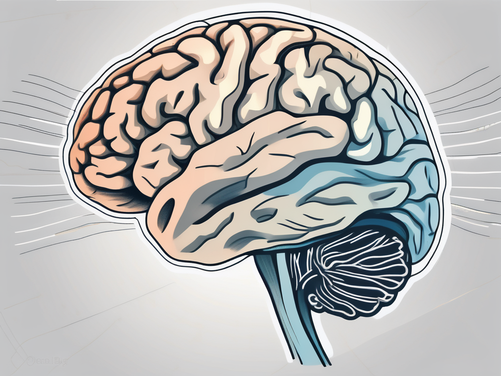
The brain, a marvel of complexity, plays a crucial role in auditory processing. Let us delve into the structure of the brain and the specific parts involved in the interpretation of sound signals.
The brain, an organ that weighs approximately three pounds, is composed of billions of interconnected neurons. It can be broadly divided into several regions, each with its own specialized functions. These regions work together seamlessly to process and interpret sensory information, including auditory signals.
The cerebral cortex, the outermost layer of the brain, is responsible for higher cognitive functions such as perception, attention, and memory. It plays a significant role in auditory perception as well. Within the cerebral cortex, specific areas, such as the auditory cortex, are dedicated to processing auditory information.
The auditory cortex, located in the temporal lobe, is responsible for processing and analyzing complex auditory information. It receives signals from the thalamus, a structure deep within the brain, and deciphers the meaning of sound. The auditory cortex is organized in a tonotopic manner, meaning that different areas within it are specialized for different frequencies of sound.
Other areas, such as the thalamus and the brainstem, also contribute to auditory processing. The thalamus acts as a relay station, directing auditory signals from the cochlea, a spiral-shaped structure in the inner ear, to the auditory cortex. It filters and amplifies the signals before they reach the cortex, ensuring that only relevant information is processed.
The brainstem, located at the base of the brain, plays a crucial role in filtering and coordinating auditory information. It receives signals from the auditory nerve, which carries information from the cochlea, and processes them before sending them to higher brain regions. The brainstem is responsible for tasks such as localizing sounds and distinguishing between different sound sources.
In addition to these specific brain regions, the brain as a whole is involved in auditory processing. It integrates auditory information with other sensory inputs and cognitive processes, allowing us to make sense of the sounds we hear and respond appropriately.
In conclusion, the brain is a remarkable organ that is intricately involved in auditory processing. From the cerebral cortex to the thalamus and brainstem, various parts work together to decipher the meaning of sound. Understanding the structure and function of these brain regions helps us appreciate the complexity of auditory perception and the wonders of the human brain.
The Pathway of Sound: From Ear to Brain
Understanding the pathway through which sound signals travel from the ear to the brain helps us comprehend the intricacies of auditory perception. Let’s embark on this sonic journey together.
As we delve deeper into the fascinating world of sound, we begin our exploration with the journey of sound waves into the ear. This journey is a remarkable process that involves various intricate mechanisms working harmoniously to enable us to perceive and interpret the sounds around us.
Journey of Sound Waves into the Ear
The journey begins when sound waves enter the outer ear and travel through the ear canal. The outer ear, also known as the pinna, acts as a funnel, directing the sound waves towards the eardrum. The eardrum, a thin, delicate membrane, starts to vibrate when struck by these sound waves, much like a drumhead responding to a beat.
From the eardrum, the vibrations are then transmitted to the middle ear, where a trio of tiny bones called the ossicles comes into play. These ossicles, consisting of the malleus (hammer), incus (anvil), and stapes (stirrup), work together as a mechanical amplifier. They amplify the vibrations received from the eardrum and transmit them to the fluid-filled cochlea, located in the inner ear.
The cochlea, often referred to as the spiral-shaped organ of hearing, is a marvel of biological engineering. Within the cochlea, the vibrations are transformed into electrical signals through a complex series of events. The vibrations cause the fluid inside the cochlea to move, stimulating thousands of microscopic hair cells lining the cochlear walls.
These hair cells, resembling delicate sensory receptors, convert the mechanical vibrations into electrical signals. This remarkable transformation is made possible by the ion channels present on the hair cell membranes. As the hair cells bend in response to the fluid movement, the ion channels open and allow ions to flow, generating electrical impulses that represent the sound waves.
Transformation of Sound Waves into Neural Signals
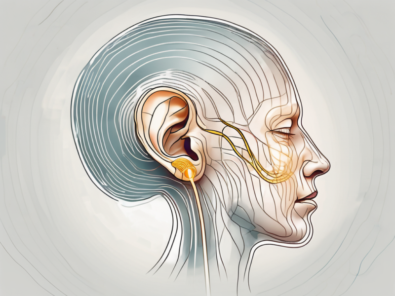
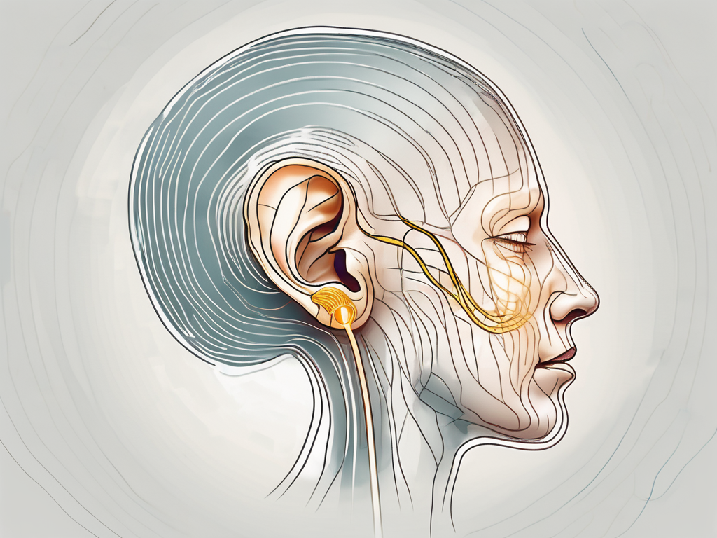
Once the cochlear nerve receives the electrical signals from the cochlea, it acts as a messenger, carrying the encoded auditory information to the brain. The cochlear nerve is a bundle of nerve fibers that extends from the cochlea and connects to the auditory centers in the brain.
Within the cochlear nerve, the auditory nerve fibers play a crucial role in relaying the electrical signals to the relevant regions of the brain. These nerve fibers, like the branches of a tree, branch out and distribute the auditory information to different areas involved in sound processing.
Upon reaching the brain, the electrical signals are received by the auditory cortex, a region responsible for sound perception and interpretation. Here, the brain performs a remarkable feat of decoding and analyzing the auditory information, allowing us to recognize and understand the sounds we hear.
The journey of sound from the ear to the brain is a complex and intricate process, involving a symphony of biological mechanisms. Understanding this pathway not only enhances our knowledge of auditory perception but also deepens our appreciation for the remarkable capabilities of the human auditory system.
The Cochlear Nerve’s Connection to the Brain
The precise mechanisms through which the cochlear nerve transmits auditory information to the brain are a subject of ongoing scientific inquiry. Nevertheless, some aspects of this connection have been elucidated.
The cochlear nerve, also known as the auditory nerve, is a crucial component of the auditory system. It is responsible for carrying electrical signals from the cochlea, the spiral-shaped structure in the inner ear, to the brain. This transmission of auditory information is essential for our ability to perceive and interpret sounds.
How the Cochlear Nerve Transmits Information
Studies suggest that the cochlear nerve transmits auditory information via a process known as spike coding. In spike coding, the electrical signals carried by the cochlear nerve are displayed as a sequence of electrical pulses, or spikes, which convey specific aspects of the auditory stimulus.
These spikes are generated by the hair cells within the cochlea. When sound waves enter the ear, they cause the hair cells to vibrate. The movement of the hair cells triggers the release of neurotransmitters, which then generate the electrical signals that travel along the cochlear nerve.
This spike code is then transmitted to the corresponding brain regions, allowing for further analysis and interpretation of the sound stimulus. The intricate timing and patterns of these spikes play a crucial role in extracting meaningful information from the auditory signal.
Destination of the Cochlear Nerve in the Brain
The cochlear nerve’s destination in the brain depends on the specific type of auditory information being processed. Different regions within the auditory cortex play a role in decoding different aspects of sound, such as pitch, timbre, and spatial location.
For example, the primary auditory cortex, located in the temporal lobe, is responsible for processing basic auditory features, such as frequency and intensity. It helps us differentiate between different pitches and loudness levels.
Moreover, the brain regions responsible for other cognitive processes, such as memory and emotion, are intertwined with the auditory processing pathways. This interconnectedness allows us to form emotional connections to sounds and recall auditory memories.
One such brain region is the amygdala, which is involved in the processing of emotions. When we hear a sound associated with a particular emotion, such as a baby’s laughter or a dog’s bark, the amygdala helps us recognize and respond to that emotion.
Another important brain region involved in auditory processing is the hippocampus, which is crucial for memory formation. It helps us remember sounds and associate them with specific events or experiences.
Overall, the connection between the cochlear nerve and the brain is a complex and intricate system that allows us to perceive, interpret, and emotionally engage with the sounds around us. Ongoing research continues to shed light on the fascinating mechanisms underlying this connection, deepening our understanding of the auditory system.
Implications of Cochlear Nerve Damage
Damage to the cochlear nerve can have profound implications on auditory perception and overall quality of life. The cochlear nerve serves as a vital bridge between the ear and the brain, enabling us to perceive and interpret the sounds that shape our world. Understanding the intricate pathways through which auditory information travels can enhance our appreciation for the remarkable complexity of the human auditory system.
When the cochlear nerve is damaged, it disrupts the normal transmission of auditory signals from the inner ear to the brain. This can result in a range of symptoms that significantly impact an individual’s ability to hear and communicate effectively.
One of the most common symptoms of cochlear nerve damage is hearing loss. This can manifest as a partial or complete inability to hear sounds, depending on the severity of the damage. Individuals may struggle to understand speech, follow conversations, or perceive certain frequencies of sound.
In addition to hearing loss, tinnitus, or ringing in the ears, is another common symptom of cochlear nerve damage. The perception of tinnitus can vary from a mild, intermittent ringing to a constant, high-pitched noise that can be extremely distressing for individuals affected by it.
Diagnosing cochlear nerve damage typically involves a comprehensive audiological evaluation. This evaluation may include a series of tests such as pure-tone audiometry, speech audiometry, and auditory brainstem response testing. These tests help assess the extent and nature of the impairment, allowing healthcare professionals to develop an appropriate treatment plan.
If you suspect you may be experiencing symptoms indicative of cochlear nerve damage, it is imperative to consult with a healthcare professional for accurate diagnosis and appropriate management strategies. Early intervention is crucial in preventing further deterioration of auditory function and improving outcomes.
Treatment and Management of Cochlear Nerve Damage
The management of cochlear nerve damage depends on the underlying cause and severity of the condition. In some cases, medical interventions, such as cochlear implants or hearing aids, may be recommended to improve auditory function and quality of life.
Cochlear implants are electronic devices that bypass the damaged cochlear nerve and directly stimulate the auditory nerve fibers. They consist of an external speech processor and an internal implant that is surgically placed in the inner ear. Cochlear implants can provide significant benefits for individuals with severe or profound hearing loss, allowing them to perceive sound and communicate more effectively.
Hearing aids, on the other hand, are amplification devices that can help individuals with mild to moderate hearing loss. They work by amplifying sound and delivering it to the ear, compensating for the impaired function of the cochlear nerve.
Additionally, audiological rehabilitation and speech therapy can provide valuable support and enhance communication skills for individuals with cochlear nerve damage. These therapies focus on improving speech perception, auditory processing, and overall communication abilities. They can also help individuals adapt to the use of hearing aids or cochlear implants, maximizing their benefits.
It is important to note that the treatment and management of cochlear nerve damage should be tailored to each individual’s specific needs and circumstances. Consulting with a healthcare professional who specializes in audiology is essential to receive the best possible care and guidance.
In conclusion, the implications of cochlear nerve damage are far-reaching and can significantly impact an individual’s auditory perception and quality of life. Understanding the symptoms, diagnosis, and treatment options for cochlear nerve damage is crucial for individuals affected by this condition. If you have any concerns or questions related to your auditory health, it is always recommended to seek guidance from a qualified medical professional to receive the best possible care.


