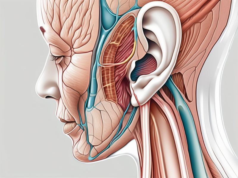
The human ear is an incredible organ that allows us to experience the world of sound. But have you ever wondered how exactly we are able to hear? In the complex process of hearing, there is one crucial action that directly stimulates nerve impulses along the cochlear nerve, and it all begins with understanding the anatomy of the ear.
Understanding the Anatomy of the Ear
The ear is a complex organ responsible for our sense of hearing. It consists of several intricate structures that work together to enable us to perceive and interpret sounds. One of these crucial structures is the cochlear nerve, which plays a vital role in the hearing process.
The Role of the Cochlear Nerve in Hearing
Before we delve into the specific action that stimulates nerve impulses, let’s first explore the role of the cochlear nerve in the hearing process. The cochlear nerve is a branch of the vestibulocochlear nerve, also known as the eighth cranial nerve. It is responsible for transmitting auditory information from the inner ear to the brain, allowing us to perceive and interpret sounds.
Imagine a symphony orchestra playing a beautiful piece of music. As the musicians play their instruments, the sound waves they produce travel through the air and enter our ears. These sound waves then pass through the various structures of the ear, including the cochlea, where the cochlear nerve resides.
The cochlear nerve acts as a messenger, carrying the information encoded in the sound waves to the brain. It is like a telephone line connecting the inner ear to the auditory processing centers in the brain. Without the cochlear nerve, our ability to hear and understand the world around us would be greatly impaired.
The Structure of the Cochlear Nerve
The cochlear nerve consists of thousands of individual nerve fibers, or axons, that originate from the hair cells within the cochlea. These hair cells are specialized sensory cells that line the organ of Corti, a structure located within the cochlea. When sound waves enter the ear, they set off a chain reaction that ultimately leads to the stimulation of these hair cells, and in turn, the cochlear nerve.
Let’s take a closer look at the structure of the cochlear nerve. Imagine it as a network of tiny cables, each carrying vital information about the sounds we hear. These cables, or axons, are incredibly thin and delicate, yet they are capable of transmitting electrical signals with remarkable precision.
Within the cochlea, the hair cells are arranged in a specific pattern, resembling the keys of a piano. Each hair cell is tuned to a specific frequency, allowing us to perceive a wide range of sounds. When sound waves enter the cochlea, they cause the fluid inside to vibrate. These vibrations, in turn, cause the hair cells to bend, generating electrical signals that travel along the axons of the cochlear nerve.
As the electrical signals travel along the cochlear nerve, they undergo a process known as neural coding. This process involves the conversion of sound waves into patterns of electrical impulses that the brain can interpret. The cochlear nerve plays a crucial role in this coding process, ensuring that the information reaches the brain accurately and efficiently.
Once the electrical signals reach the brain, they are processed and interpreted, allowing us to perceive and understand the sounds we hear. This intricate system of the cochlear nerve and its associated structures is a testament to the complexity and beauty of the human auditory system.
The Process of Hearing Explained
How Sound Waves are Transformed into Nerve Impulses
Now that we understand the basic structure of the cochlear nerve, let’s explore how sound waves are transformed into nerve impulses. The journey begins when sound waves enter the ear canal and reach the eardrum. The eardrum, also known as the tympanic membrane, is a thin, cone-shaped layer of tissue that separates the outer ear from the middle ear. It vibrates in response to the sound waves, much like a drumhead, amplifying the sound and transmitting it further into the ear.
As the eardrum vibrates, it sets into motion the tiny bones of the middle ear – the ossicles. The ossicles consist of three bones: the malleus, incus, and stapes. These bones work together as a lever system, amplifying the sound vibrations and transmitting them to the fluid-filled cochlea. The malleus, also known as the hammer, is attached to the eardrum and receives the vibrations. It then transfers these vibrations to the incus, or anvil, which in turn passes them on to the stapes, or stirrup. The stapes, the smallest bone in the human body, acts as a piston, pushing against the oval window of the cochlea.
Within the cochlea, the sound vibrations cause the fluid to move, stimulating the hair cells. The cochlea is a spiral-shaped, snail-like structure that is filled with fluid. It is divided into three fluid-filled compartments: the scala vestibuli, the scala media, and the scala tympani. The scala media, also known as the cochlear duct, is where the magic happens. It contains the organ of Corti, which is responsible for converting sound vibrations into electrical signals.
The organ of Corti is a complex structure that contains thousands of hair cells. These hair cells are equipped with tiny hair-like projections called stereocilia. The stereocilia are arranged in rows and are graded in height, with the tallest ones located at the outer edge and the shortest ones at the inner edge. When the sound vibrations cause the fluid in the cochlea to move, the stereocilia bend in response to the fluid movement.
When the stereocilia bend, ion channels open, allowing electrically charged particles to enter the hair cells. This generates an electrical signal that triggers the release of neurotransmitters, signaling the nearby cochlear nerve fibers to transmit the auditory information to the brain. The cochlear nerve fibers are part of the auditory nerve, which is responsible for carrying sound information from the cochlea to the brain.
The Journey of Sound: From Ear Canal to Cochlear Nerve
Let’s take a moment to appreciate the intricate journey that sound takes from the ear canal to the cochlear nerve. After the electrical signal is generated in the hair cells, it travels along the cochlear nerve fibers towards the brain. These fibers are organized tonotopically, meaning they transmit information about specific frequencies of sound.
As the auditory information reaches the brain, it passes through various processing centers where it is interpreted and integrated. The primary auditory cortex, located in the temporal lobe of the brain, is responsible for processing basic sound information, such as pitch and loudness. From there, the processed information is sent to other areas of the brain, such as the auditory association cortex, where higher-level processing takes place.
The brain analyzes the different frequencies, loudness, and timing of the sounds, allowing us to perceive and recognize the world of auditory stimuli around us. It is through this complex process that we are able to enjoy music, understand speech, and navigate our environment using sound cues.
The Action that Stimulates Nerve Impulses
When it comes to stimulating nerve impulses along the cochlear nerve, there is a specific action that plays a crucial role. It is the bending of the stereocilia within the hair cells. These hair cells are located in the inner ear and are responsible for converting sound vibrations into electrical signals that can be interpreted by the brain.
So, how does this process work? When sound vibrations enter the ear, they cause fluid movement in the cochlea, a spiral-shaped structure in the inner ear. This fluid movement, in turn, leads to the displacement of the stereocilia. As the fluid moves, it pushes against the hair cells, causing the stereocilia to bend.
It is this mechanical stimulation of the hair cells that triggers the opening of ion channels. These ion channels are tiny pores located on the surface of the hair cells. When the stereocilia bend, these ion channels open up, allowing ions to flow in and out of the hair cells.
As ions flow in and out, electrical signals are generated within the hair cells. These electrical signals, also known as action potentials, are the language of the nervous system. They are the means by which information is transmitted from the ear to the brain.
Once the electrical signals are generated, they are transmitted to the cochlear nerve. This nerve carries the signals from the hair cells to the brain, where they are interpreted as sound. In this way, the bending of the stereocilia within the hair cells is the crucial action that directly stimulates nerve impulses along the cochlear nerve.
The Role of Hair Cells in Stimulating Nerve Impulses
Now that we understand how the bending of the stereocilia stimulates nerve impulses, let’s take a closer look at the role of hair cells in this process. Hair cells are specialized sensory cells that are found in the inner ear. They are named after the hair-like structures, called stereocilia, that protrude from their surface.
These hair cells are arranged in rows within the cochlea, with each row having a specific function. The outer hair cells, for example, are responsible for amplifying sound signals, while the inner hair cells are responsible for converting sound vibrations into electrical signals.
When sound waves enter the ear, they cause the fluid in the cochlea to move. This fluid movement, in turn, causes the stereocilia to bend. The bending of the stereocilia is what triggers the hair cells to generate electrical signals.
It is important to note that hair cells are highly specialized and delicate structures. They are sensitive to even the slightest movements and can be easily damaged. This is why exposure to loud noises over a prolonged period of time can lead to hearing loss.
In addition to their role in stimulating nerve impulses, hair cells also play a crucial role in the perception of pitch and loudness. The arrangement and sensitivity of the hair cells allow us to distinguish between different frequencies of sound and perceive differences in volume.
The Impact of Sound Intensity on Nerve Impulse Stimulation
While we have discussed how the bending of stereocilia stimulates nerve impulses, it is worth noting that the intensity of sound also plays a crucial role in this process. Sound intensity refers to the loudness or strength of a sound.
When a sound is more intense, or louder, it causes a greater displacement of the stereocilia. This means that the hair cells are bent to a greater degree, resulting in a stronger electrical signal being generated.
So, why does this matter? The greater the displacement of the stereocilia and the stronger the electrical signal generated, the more intense the nerve impulse that is transmitted to the brain. This allows us to perceive differences in volume and loudness in the sounds we hear.
For example, when we listen to a loud concert, the intense sound vibrations cause a significant displacement of the stereocilia. This results in a strong electrical signal being generated by the hair cells, which is then transmitted to the brain. As a result, we perceive the sound as being loud.
In contrast, when we listen to a soft whisper, the sound vibrations are less intense, causing a smaller displacement of the stereocilia. This leads to a weaker electrical signal being generated by the hair cells, which is then transmitted to the brain. Consequently, we perceive the sound as being soft.
Therefore, the impact of sound intensity on nerve impulse stimulation is crucial in our ability to perceive and interpret the sounds around us.
The Connection Between Cochlear Nerve and Brain
The cochlear nerve plays a crucial role in our ability to hear and interpret sounds. This intricate connection between the cochlear nerve and the brain allows us to have a rich auditory experience and navigate the world through sound.
How Nerve Impulses are Interpreted by the Brain
Once the nerve impulses are transmitted along the cochlear nerve to the brain, they embark on a fascinating journey of interpretation and processing. The brain, being the master conductor of our auditory system, meticulously decodes the frequency, intensity, and timing of these nerve impulses.
Imagine the brain as a skilled orchestra conductor, carefully analyzing the incoming signals to create a symphony of sound perception. It dissects the nerve impulses, extracting valuable information that allows us to perceive different pitches, loudness levels, and even the location of sounds in our environment.
This complex process is what enables us to recognize speech, enjoy the melodies of music, and be fully aware of our surroundings through sound. It is through the intricate interpretation of these nerve impulses that we are able to appreciate the beauty and significance of the auditory world.
The Cochlear Nerve’s Role in Transmitting Sound Information to the Brain
Without the cochlear nerve, the transmission of sound information from the inner ear to the brain would be impossible. This nerve acts as the vital link in the auditory pathway, ensuring that the electrical signals generated by the hair cells in the cochlea reach the appropriate centers within the brain for processing and interpretation.
Think of the cochlear nerve as a reliable messenger, faithfully delivering the important auditory messages from the inner ear to the brain. It acts as a conduit, allowing the electrical impulses to travel along its intricate fibers, ultimately reaching the brain’s auditory centers.
These fibers of the cochlear nerve are finely tuned to carry specific information related to sound. Each fiber is responsible for transmitting a particular frequency or pitch, ensuring that the brain receives a comprehensive representation of the entire auditory spectrum.
As the nerve impulses travel along the cochlear nerve, they encounter various checkpoints and relay stations, where they are carefully monitored and modulated. This ensures that the information is accurately transmitted and that any potential distortions or disruptions are minimized.
Once the nerve impulses successfully reach the brain, they are met with a complex network of neurons and intricate neural pathways. These pathways work in harmony to process, interpret, and integrate the incoming auditory information, ultimately allowing us to perceive and make sense of the sounds around us.
So, the connection between the cochlear nerve and the brain is not just a simple transmission of electrical signals. It is a remarkable collaboration between the delicate structures of the inner ear and the intricate neural networks of the brain, working together to give us the gift of hearing.
Disorders Related to the Cochlear Nerve
Symptoms and Causes of Cochlear Nerve Damage
While the cochlear nerve is a remarkable part of our auditory system, it can sometimes be affected by disorders that lead to hearing impairment. Damage to the cochlear nerve can result from various conditions, including noise-induced hearing loss, infections, tumors, or genetic factors. Symptoms of cochlear nerve damage may include hearing loss, tinnitus, dizziness, or difficulty understanding speech.
If you experience any of these symptoms, it is vital to consult with a medical professional, such as an otolaryngologist or audiologist, for a proper evaluation and diagnosis. A thorough assessment can help determine the underlying cause of the symptoms and guide appropriate treatment options.
Treatment and Prevention of Cochlear Nerve Disorders
The treatment and prevention of cochlear nerve disorders depend on the specific underlying cause. In some cases, medical interventions, such as surgeries or hearing aids, may be recommended. Advanced technologies, like cochlear implants, can also be beneficial for individuals with severe or profound hearing loss resulting from cochlear nerve damage.
Prevention strategies for cochlear nerve disorders include protecting our hearing from loud noises, practicing good ear hygiene, and seeking prompt medical attention for any ear-related symptoms or infections. Regular hearing screenings and check-ups can help detect any potential issues early on and allow for timely intervention.
Conclusion
In the process of hearing, it is the action of the bending stereocilia within the hair cells that directly stimulates nerve impulses along the cochlear nerve. This action sets in motion a complex chain of events that ultimately allows us to experience the richness of sound. Understanding the intricate anatomy of the ear and the role of the cochlear nerve provides us with a deeper appreciation of the remarkable mechanisms that enable the gift of hearing. Should you encounter any concerns about your hearing or experience symptoms related to the cochlear nerve, always seek professional medical advice to ensure an accurate diagnosis and appropriate treatment.


