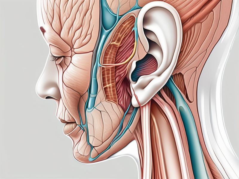
he cochlear nerve is a crucial component of our auditory system, responsible for transmitting sound information from the inner ear to the brain. Understanding the intricacies of this nerve can help shed light on the fascinating world of hearing and its underlying mechanisms. In this article, we will explore the anatomy and function of the cochlear nerve, delve into the world of neurons within this nerve, examine the methods for counting them, and discuss the significance of neuron count in relation to hearing abilities and neurological disorders. Additionally, we will address some frequently asked questions to provide a comprehensive understanding of the topic.
Understanding the Cochlear Nerve
The cochlear nerve is a crucial component of the auditory nerve complex, responsible for the transmission of auditory signals. It is a bilateral nerve, meaning that it exists in both ears and connects the inner ear to the brainstem.
The cochlear nerve originates from the spiral ganglion, a cluster of specialized nerve cells located in the cochlea of the inner ear. These nerve cells play a vital role in converting sound waves into electrical signals that can be interpreted by the brain. From the spiral ganglion, the cochlear nerve travels through the internal auditory canal, a narrow passageway within the temporal bone.
Upon reaching the brainstem, the cochlear nerve divides into two main branches: the vestibular division and the cochlear division. While the vestibular division is responsible for transmitting information related to balance and spatial orientation, the cochlear division is primarily dedicated to carrying auditory signals.
Anatomy of the Cochlear Nerve
The cochlear nerve consists of thousands of individual nerve fibers, each playing a crucial role in the transmission of sound information. These fibers are organized in a complex manner, with different regions of the cochlea corresponding to specific frequencies of sound.
Within the cochlea, there are three fluid-filled chambers known as the scala vestibuli, the scala media, and the scala tympani. The scala media, also known as the cochlear duct, contains the organ of Corti, which houses the sensory hair cells responsible for detecting sound vibrations.
When sound waves enter the ear canal and reach the cochlea, they cause the fluid within the scala vestibuli to vibrate. These vibrations are then transmitted to the scala media, causing the sensory hair cells to bend. The bending of these hair cells triggers the release of neurotransmitters, which in turn generate electrical signals that can be picked up by the nerve fibers of the cochlear nerve.
Function of the Cochlear Nerve
The primary function of the cochlear nerve is to transmit auditory information from the inner ear to the brain. Once the electrical signals generated by the hair cells reach the cochlear nerve, they are carried to the brainstem, specifically the cochlear nucleus.
Within the cochlear nucleus, the auditory signals undergo further processing and interpretation. Different regions of the cochlear nucleus are responsible for analyzing specific aspects of sound, such as pitch, intensity, and timing. This complex processing allows the brain to perceive and understand the various qualities of sound.
From the cochlear nucleus, the auditory signals are relayed to higher brain regions, including the inferior colliculus and the auditory cortex. These regions are involved in more advanced processing and integration of auditory information, ultimately leading to the perception of sound.
It is important to note that the cochlear nerve not only transmits sound information but also plays a role in the regulation of hearing sensitivity. The nerve fibers within the cochlear nerve can adjust their sensitivity to different sound levels, allowing for optimal hearing in various environments.
In summary, the cochlear nerve is a vital component of the auditory system, responsible for transmitting auditory signals from the inner ear to the brain. Its intricate anatomy and complex processing mechanisms enable us to perceive and understand the rich world of sound that surrounds us.
The Neurons in the Cochlear Nerve
What are Neurons?
Neurons, often referred to as nerve cells, are the basic building blocks of the nervous system. These specialized cells are responsible for transmitting electrical signals and information throughout the body. They are highly complex and have unique structures that enable them to carry out their functions effectively. Neurons consist of three main parts: the cell body, dendrites, and axon.
The cell body, also known as the soma, contains the nucleus and other organelles necessary for the neuron’s survival and functioning. It processes incoming signals and generates electrical impulses. Dendrites, on the other hand, are branch-like structures that extend from the cell body and receive signals from other neurons or sensory receptors. They play a crucial role in integrating and transmitting information towards the cell body.
The axon is a long, slender projection that carries electrical impulses away from the cell body and towards other neurons or target cells. It is covered by a protective layer called the myelin sheath, which enhances the speed of signal transmission. At the end of the axon, there are small branches called axon terminals, which form connections with other neurons or target cells.
In the context of the cochlear nerve, neurons play a vital role in transmitting auditory information from the inner ear to the brainstem for further processing. This intricate process involves the conversion of sound waves into electrical signals that can be interpreted by the brain.
Role of Neurons in the Cochlear Nerve
Within the cochlear nerve, neurons serve as the communicators, transmitting electrical signals generated by the hair cells in the cochlea to the brainstem. These signals are crucial for our perception of sound. The cochlea, a spiral-shaped structure in the inner ear, contains thousands of hair cells that convert sound vibrations into electrical signals.
Once the hair cells detect sound waves, they release neurotransmitters that stimulate the nearby neurons in the cochlear nerve. These neurons then carry the electrical signals towards the brainstem, where further processing and interpretation of the auditory information occur.
Neurons in the cochlear nerve form numerous connections, allowing for efficient transmission and integration of auditory information. They are organized in a precise manner, with different groups of neurons responsible for transmitting specific frequencies of sound. This organization enables us to perceive and differentiate various pitches and tones.
Furthermore, the neurons in the cochlear nerve play a crucial role in maintaining the balance between sensitivity and selectivity in our auditory system. They help filter out irrelevant background noise and focus on the sounds that are important for our perception and understanding of the environment.
In conclusion, the neurons in the cochlear nerve are essential for the transmission of auditory information from the inner ear to the brainstem. Their intricate structure and precise organization enable efficient signal transmission and integration, allowing us to perceive and interpret the sounds around us.
Counting the Neurons in the Cochlear Nerve
The cochlear nerve, a vital component of the auditory system, plays a crucial role in transmitting auditory information from the inner ear to the brain. Understanding the exact number of neurons present in the cochlear nerve is a challenging task due to its complex and intricate structure.
Methods for Counting Neurons
Researchers have developed various methods to estimate the neuron count in the cochlear nerve. These methods involve a combination of histological analysis and stereology.
Histological analysis is a technique that involves the examination of tissue samples under a microscope. By carefully studying these samples, researchers can identify and count the neurons present in the cochlear nerve. This method provides valuable insights into the cellular composition of the nerve and allows for a more accurate estimation of neuron count.
Another method used to estimate neuron count is stereology. Stereology utilizes mathematical principles to extrapolate the total number of neurons from sampled areas of the cochlear nerve. By systematically analyzing these samples, researchers can make reliable predictions about the overall neuron count in the nerve.
Estimated Number of Neurons in the Cochlear Nerve
The estimated number of neurons in the cochlear nerve can vary depending on the study and the methodology used. However, current research suggests that the human cochlear nerve is composed of thousands of individual neurons. This intricate network of neurons ensures the efficient transmission of auditory information from the inner ear to the brain.
It is important to note that individual variations in neuron count may exist among individuals. Factors such as age, genetics, and environmental influences can contribute to these variations. Therefore, while estimates provide a general understanding of neuron count in the cochlear nerve, it is essential to consider the potential for individual differences.
Further research is ongoing to refine the methods used for counting neurons in the cochlear nerve. By improving our understanding of the cellular composition of this vital nerve, researchers can gain valuable insights into the mechanisms underlying hearing and develop more effective treatments for auditory disorders.
Significance of Neuron Count in the Cochlear Nerve
The cochlear nerve plays a crucial role in our ability to hear, and the number of neurons within this nerve has significant implications for our hearing abilities. The cochlear nerve is responsible for transmitting auditory signals from the inner ear to the brain, where they are processed and interpreted as sound. If the number of neurons in the cochlear nerve is compromised, either due to developmental abnormalities or damage, it can lead to a reduction in the efficiency of auditory signal transmission.
Imagine a complex network of highways connecting different cities. Each neuron within the cochlear nerve acts as a messenger, carrying important information about sound from the ear to the brain. When there is a sufficient number of neurons, this network functions smoothly, allowing for clear and accurate transmission of auditory signals. However, when the neuron count is reduced, it’s like having fewer messengers on the road, causing delays and disruptions in the delivery of sound information.
This reduction in the efficiency of auditory signal transmission can manifest as hearing loss or difficulties in perceiving certain sounds. People with a lower neuron count in the cochlear nerve may struggle to hear soft sounds, have trouble understanding speech in noisy environments, or experience a decreased ability to discriminate between different pitches or frequencies.
It is important to note that while neuron count in the cochlear nerve is an important factor in hearing abilities, it is just one piece of the puzzle. Other factors, such as the health and integrity of the hair cells in the inner ear, the function of the auditory pathways in the brain, and the overall cognitive processing of sound, also contribute to our ability to hear and understand the world around us.
Implications for Neurological Disorders
Understanding the relationship between neuron count in the cochlear nerve and neurological disorders is an area of active research. Researchers are investigating how abnormalities in the cochlear nerve and its neuron count may contribute to various neurological conditions, such as tinnitus or auditory neuropathy.
Tinnitus, often described as a persistent ringing or buzzing sound in the ears, is a common condition that can significantly impact a person’s quality of life. While the exact cause of tinnitus is still not fully understood, some studies have suggested that it may be associated with abnormalities in the cochlear nerve. It is believed that changes in the neuron count or function within the cochlear nerve may contribute to the perception of phantom sounds, leading to the sensation of tinnitus.
Auditory neuropathy is another neurological disorder that has been linked to abnormalities in the cochlear nerve. This condition is characterized by a disconnect between the transmission of auditory signals from the inner ear to the brain. Individuals with auditory neuropathy may have difficulty understanding speech, despite having relatively normal hearing thresholds. It is thought that disruptions in the neuron count or function within the cochlear nerve may contribute to this disorder.
However, it is important to note that the relationship between neuron count in the cochlear nerve and these neurological disorders is complex and multifaceted. Further research is needed to fully elucidate the mechanisms underlying these conditions and to develop effective treatments that target the specific abnormalities within the cochlear nerve.
Frequently Asked Questions about the Cochlear Nerve and Neurons
Can the Number of Neurons Change?
The number of neurons in the cochlear nerve is generally established during development and remains relatively constant throughout adulthood. However, it is important to note that certain factors, such as age-related degeneration or specific medical conditions, may lead to a decrease in neuron count.
Age-related degeneration is a natural process that occurs as we grow older. Over time, the cochlear nerve may experience some degeneration, which can result in a slight decrease in the number of neurons. This degeneration can be influenced by various factors, including genetics, exposure to loud noises, and overall health.
Additionally, specific medical conditions, such as certain types of tumors or infections, can also affect the number of neurons in the cochlear nerve. These conditions may cause damage or destruction to the nerve cells, leading to a reduction in neuron count.
Consultation with a healthcare professional, such as an otolaryngologist or audiologist, can provide a more personalized evaluation and guidance in these situations. They can assess your individual circumstances and provide recommendations on how to manage any changes in neuron count that may occur.
What Happens if Neurons in the Cochlear Nerve are Damaged?
If neurons in the cochlear nerve are damaged, either due to injury, disease, or other factors, it can result in hearing loss or auditory dysfunction. The extent and impact of such damage can vary depending on the severity and location of the injury.
When neurons in the cochlear nerve are damaged, the transmission of auditory information from the inner ear to the brainstem is disrupted. This can lead to difficulties in perceiving and understanding sounds, as well as challenges in distinguishing between different frequencies and volumes.
There are various causes of cochlear nerve damage, including exposure to loud noises, certain medications, infections, and head trauma. In some cases, the damage may be reversible with appropriate medical intervention, while in others, it may be permanent.
If you are experiencing any changes in your hearing abilities or have concerns regarding your auditory health, it is advisable to seek a consultation with a medical professional who specializes in ear and hearing disorders. They can conduct a thorough evaluation, including hearing tests and imaging studies, to determine the extent of the damage and recommend appropriate treatment options.
It is important to address any issues with the cochlear nerve promptly, as untreated damage can have a significant impact on an individual’s quality of life. With advancements in medical technology and rehabilitation techniques, there are often effective strategies available to manage and improve hearing function.
In conclusion, the cochlear nerve, with its intricate anatomy and vital role in sound transmission, is an essential component of our auditory system. Neurons within this nerve facilitate the efficient transfer of auditory information from the inner ear to the brainstem. Understanding the neuron count in the cochlear nerve can shed light on hearing abilities and may have implications for the diagnosis and management of certain neurological disorders.
However, it is important to remember that professional evaluation and guidance from healthcare experts are crucial for addressing individual concerns and queries related to the cochlear nerve and its neurons. Each person’s situation is unique, and a comprehensive assessment by a qualified medical professional is necessary to provide accurate information and personalized recommendations.


