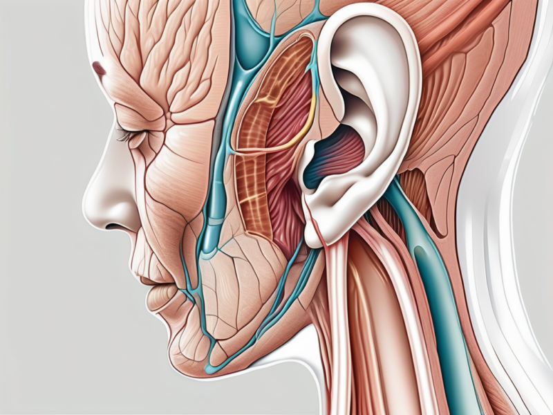
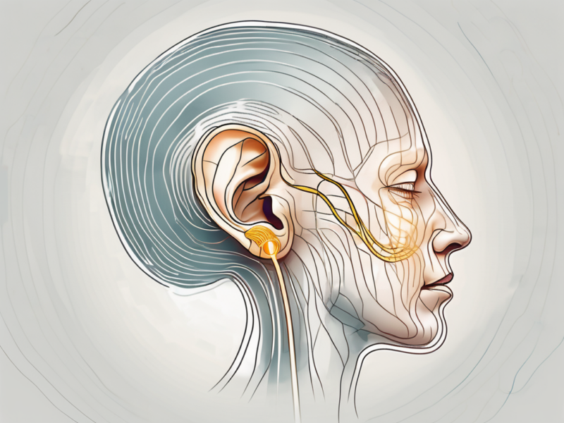
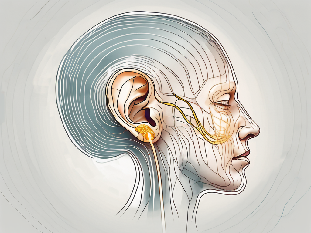
The cochlear nerve is an essential component of the auditory system, playing a crucial role in our ability to hear and perceive sound. To fully understand the function of the cochlear nerve, it is vital to delve into the complex anatomy of the ear and explore its interconnections with other structures.
Understanding the Anatomy of the Ear
The ear is a complex organ responsible for our ability to hear and perceive sound. It is composed of several intricate structures, each playing a crucial role in the auditory process. One of these structures is the cochlear nerve, which is located within the inner ear.
The Role of the Cochlear Nerve in the Ear
The cochlear nerve is a branch of the vestibulocochlear nerve, specifically dedicated to transmitting auditory information from the cochlea to the brain. Without this nerve, our ability to hear and interpret sound would be greatly impaired.
When sound waves enter the ear, they travel through the ear canal and reach the eardrum. The eardrum vibrates in response to these sound waves, causing the tiny bones in the middle ear to move. These movements amplify the sound and transmit it to the cochlea, a spiral-shaped structure within the inner ear.
Within the cochlea, the cochlear nerve plays a vital role in converting these mechanical vibrations into electrical signals that can be interpreted by the brain. It is a sensory nerve that carries electrical signals known as action potentials, enabling us to perceive sound.
The Structure of the Cochlear Nerve
The cochlear nerve is composed of approximately 30,000 individual sensory fibers that originate from the spiral ganglion, a cluster of nerve cell bodies within the cochlea. These fibers form a complex network and travel through the bony cochlear canal, eventually joining together to form the cochlear nerve bundle.
As the cochlear nerve fibers travel through the cochlear canal, they are organized according to the specific frequencies of sound they respond to. This organization allows for the precise encoding and transmission of different pitches and tones to the brain.
Once the cochlear nerve fibers reach the brain, they synapse with neurons in the auditory cortex, where the electrical signals are further processed and interpreted as sound. This intricate network of nerve fibers and synapses ensures that we can perceive and understand the wide range of sounds in our environment.
In conclusion, the cochlear nerve is a vital component of the ear’s anatomy, responsible for transmitting auditory information from the cochlea to the brain. Its complex structure and organization allow for the precise encoding and interpretation of sound, enabling us to enjoy the rich and diverse world of auditory experiences.
The Function of the Cochlear Nerve
Sound Transmission Process
When sound waves enter the ear, they cause vibrations of the air molecules. These vibrations are then transmitted through the ear canal and reach the eardrum, causing it to vibrate as well. The eardrum’s vibrations are essential for the process of hearing.
As the vibrations travel through the ear, they encounter the middle ear, where the ossicles, a set of tiny bones, amplify the sound. The ossicles consist of the malleus, incus, and stapes, which work together to transmit the vibrations from the eardrum to the cochlea.
The cochlea, a spiral-shaped, fluid-filled structure, is the main sensory organ responsible for hearing. It is divided into three fluid-filled compartments: the scala vestibuli, the scala media, and the scala tympani. These compartments are separated by membranes and contain different structures that play crucial roles in the hearing process.
Within the cochlea, the sound waves create ripples in the fluid, stimulating thousands of hair cells lining the organ of Corti. These hair cells are the sensory receptors responsible for converting the mechanical vibrations into electrical signals, which can be interpreted by the brain as sound.
The electrical signals generated by the hair cells are picked up by the delicate fibers of the cochlear nerve. This nerve carries the auditory information from the cochlea to the brain, where it is processed and interpreted as sound.
The Cochlear Nerve and Balance
Although primarily responsible for hearing, the cochlear nerve also plays a role in maintaining balance. In addition to receiving auditory information, the cochlear nerve receives input from specialized sensory cells in the vestibular system.
The vestibular system is responsible for detecting changes in head position and movement, providing crucial information for coordinating our body’s balance and spatial orientation. The sensory cells in the vestibular system detect the movement of fluid within the semicircular canals of the inner ear, which are responsible for detecting rotational movements.
By receiving information from the vestibular system, the cochlear nerve contributes to our ability to maintain balance and equilibrium. It helps us adjust our body position and make precise movements, allowing us to navigate our surroundings with ease.
Disorders Related to the Cochlear Nerve
The cochlear nerve is a vital component of the auditory system, responsible for transmitting sound signals from the inner ear to the brain. However, like any other part of the body, it can be susceptible to damage or dysfunction, leading to various disorders.
Symptoms of Cochlear Nerve Damage
When the cochlear nerve is affected, individuals may experience a range of symptoms that can significantly impact their quality of life. One of the most common signs is hearing loss, which can vary in severity from mild to profound. This hearing impairment can make it challenging to understand speech, particularly in noisy environments.
In addition to hearing loss, those with cochlear nerve damage may also experience tinnitus, a persistent ringing, buzzing, or hissing sound in the ears. This phantom noise can be incredibly bothersome and may interfere with daily activities and concentration.
Furthermore, some individuals may encounter issues with balance and coordination due to the connection between the cochlear nerve and the vestibular system, responsible for maintaining equilibrium. Imbalance problems can lead to dizziness, vertigo, and a feeling of unsteadiness.
Recognizing these symptoms is crucial, as early intervention can significantly improve outcomes. If you or someone you know is experiencing any of these signs, it is essential to consult with a qualified healthcare professional for an accurate diagnosis.
Diagnosis and Treatment Options
When evaluating suspected cochlear nerve disorders, doctors employ a comprehensive approach to determine the underlying cause and develop an appropriate treatment plan. The diagnostic process typically involves a series of tests and assessments.
A comprehensive hearing assessment is one of the primary tools used to evaluate cochlear nerve function. This evaluation may include pure-tone audiometry, speech audiometry, and tympanometry. These tests help determine the extent and nature of the hearing loss and provide valuable information for treatment planning.
In some cases, additional imaging studies may be necessary to visualize the cochlear nerve and surrounding structures. Magnetic resonance imaging (MRI) or computed tomography (CT) scans can provide detailed images of the inner ear, helping identify any structural abnormalities or lesions that may be affecting the cochlear nerve.
Once a diagnosis has been established, treatment options can be explored. The specific approach will depend on the underlying condition and its severity. For individuals with mild to moderate hearing loss, hearing aids may be recommended. These devices amplify sound, making it easier for the cochlear nerve to transmit signals to the brain.
In cases of severe or profound hearing loss, cochlear implants may be considered. These electronic devices bypass the damaged cochlear nerve and directly stimulate the auditory nerve, allowing individuals to perceive sound. Cochlear implants can significantly improve hearing ability and speech comprehension in suitable candidates.
Additionally, management strategies for cochlear nerve disorders may involve medical interventions or therapy. Medications can be prescribed to address specific underlying causes, such as inflammation or infection. Rehabilitation programs, including auditory training and speech therapy, can also play a vital role in maximizing communication skills and adapting to hearing loss.
It is important to remember that each individual’s situation is unique, and treatment plans should be tailored to their specific needs. Regular follow-up appointments with healthcare professionals are essential to monitor progress and make any necessary adjustments to the treatment approach.
The Cochlear Nerve and Hearing Loss
Cochlear Implants: How They Work
For individuals with severe to profound hearing loss, cochlear implants can be a life-changing solution. These devices bypass the damaged cochlear nerve by directly stimulating the auditory nerve fibers. A surgically implanted component captures sound signals, converts them into electrical impulses, and sends them to the acoustic nerve, allowing individuals to perceive sound.
Let’s delve deeper into how cochlear implants work. The surgically implanted component consists of two main parts: an external microphone and speech processor, and an internal receiver and electrode array. The external microphone picks up sounds from the environment and sends them to the speech processor, which analyzes and converts the sounds into digital signals.
Once the sounds are converted into digital signals, they are sent to the internal receiver and electrode array. The receiver, which is placed under the skin behind the ear, receives the signals and transmits them to the electrode array. The electrode array, which is inserted into the cochlea, stimulates the auditory nerve fibers directly, bypassing the damaged cochlear nerve.
Each electrode in the array corresponds to a specific frequency range, allowing for a more detailed representation of sound. When the electrical impulses reach the auditory nerve fibers, they are sent to the brain, where they are interpreted as sound. With the help of cochlear implants, individuals with severe to profound hearing loss can regain their ability to perceive sound and improve their overall quality of life.
The Impact of Aging on the Cochlear Nerve
The aging process can affect the cochlear nerve and lead to age-related hearing loss, known as presbycusis. Over time, the nerve fibers may degenerate, reducing the transmission of auditory signals to the brain. While age-related hearing loss is a natural part of the aging process, seeking professional advice can help identify strategies for managing and adapting to these changes.
Presbycusis typically starts with a gradual decline in the ability to hear high-frequency sounds. As the cochlear nerve degenerates, it becomes more challenging to understand speech, especially in noisy environments. This can lead to difficulties in communication and social interactions, impacting an individual’s overall well-being.
It is important to note that age-related hearing loss is not solely caused by the degeneration of the cochlear nerve. Other factors, such as exposure to loud noises throughout life, genetics, and certain medical conditions, can also contribute to the development of presbycusis.
While there is no cure for age-related hearing loss, various interventions can help manage its impact. Hearing aids, for example, can amplify sounds and improve speech understanding. Assistive listening devices, such as FM systems or captioned telephones, can also be beneficial in specific situations.
Additionally, speech and hearing professionals can provide counseling and communication strategies to help individuals cope with the challenges of age-related hearing loss. These strategies may include techniques for effective communication, such as facing the person directly, speaking clearly, and minimizing background noise.
Regular hearing evaluations are essential for monitoring changes in hearing abilities and adjusting interventions accordingly. By staying proactive and seeking professional advice, individuals with age-related hearing loss can continue to engage in their daily activities and maintain a high quality of life.
The Future of Cochlear Nerve Research
Advances in Cochlear Nerve Regeneration
Researchers are continually exploring new avenues to enhance the function of the cochlear nerve. One area of focus is regenerating damaged nerve fibers, aiming to restore normal hearing and provide hope for individuals with hearing loss caused by nerve damage. Although these advancements are still in the experimental stage, they hold great promise for the future.
Recent studies have shown that certain growth factors, such as brain-derived neurotrophic factor (BDNF) and glial cell line-derived neurotrophic factor (GDNF), have the potential to stimulate the growth of new nerve fibers in the cochlea. By delivering these growth factors directly to the damaged areas, researchers hope to promote the regeneration of the cochlear nerve and improve hearing outcomes.
Furthermore, researchers are investigating the use of gene therapy to enhance the regeneration process. By introducing specific genes into the damaged cells, scientists aim to activate the natural regenerative capabilities of the cochlear nerve. This approach shows promise in preclinical studies and may offer a new avenue for treating hearing loss caused by nerve damage.
The Potential of Stem Cell Therapy
Another exciting area of research is the use of stem cells to repair and regenerate damaged cochlear nerve fibers. Stem cells have the potential to develop into various cell types, including nerve cells. By introducing stem cells into the damaged areas, scientists aim to promote the growth of new nerve fibers, leading to improved hearing outcomes. However, further studies and clinical trials are needed to fully understand the safety and efficacy of this approach.
Researchers are also exploring different sources of stem cells for cochlear nerve regeneration. While embryonic stem cells have shown promise in early studies, ethical concerns and technical challenges limit their widespread use. As an alternative, induced pluripotent stem cells (iPSCs) derived from adult cells offer a promising solution. These iPSCs can be reprogrammed to behave like embryonic stem cells, providing a potentially unlimited source of cells for regenerative therapies.
In addition to stem cell transplantation, scientists are investigating the use of biomaterials to create a supportive environment for nerve regeneration. These biomaterials can provide structural support, release growth factors, and guide the growth of new nerve fibers. By combining stem cell therapy with biomaterials, researchers hope to optimize the regenerative process and improve hearing outcomes for individuals with cochlear nerve damage.
In conclusion, the cochlear nerve plays a vital role in our auditory system, allowing us to perceive sound and maintain balance. Understanding the complex anatomy of the ear and the interconnections between its various components is crucial for comprehending the function of the cochlear nerve. While disorders related to the cochlear nerve can have a significant impact on an individual’s quality of life, advancements in research and treatment options offer hope for improving hearing outcomes. If you have concerns regarding the function of your cochlear nerve or experience any symptoms related to hearing loss, it is important to consult with a medical professional who specializes in ear health and hearing disorders.


